Cone Beam 3D (CBCT)
A three-dimensional image of your teeth and jaw, to pinpoint to the millimetre what a conventional X-ray does not show. It is useful before an implant or a complex root canal treatment. Painless, in a few seconds.

CID (Cabinet d’Imagerie Dentaire)
Dental imaging by Dubois
Dental imaging facilities in Luxembourg-Merl, by referral only from a dentist, an orthodontist or a surgeon. A clinical report is sent to the referring practitioner within 24 to 48 working hours.
The Cabinet d’Imagerie Dentaire (CID) brings together in Luxembourg-Merl the three examinations used in dental diagnosis: Cone Beam 3D, the panoramic X-ray and the lateral cephalogram. Our equipment is digital and the field of view is limited to the area referred, which reduces the dose delivered compared with a wide-field examination.
All examinations are carried out by referral only. This is not an administrative formality: the clinical indication given by the referring practitioner determines the area examined, and therefore the exposure. The images are interpreted and then sent with a written clinical report within 24 to 48 working hours. You have nothing to arrange yourself.
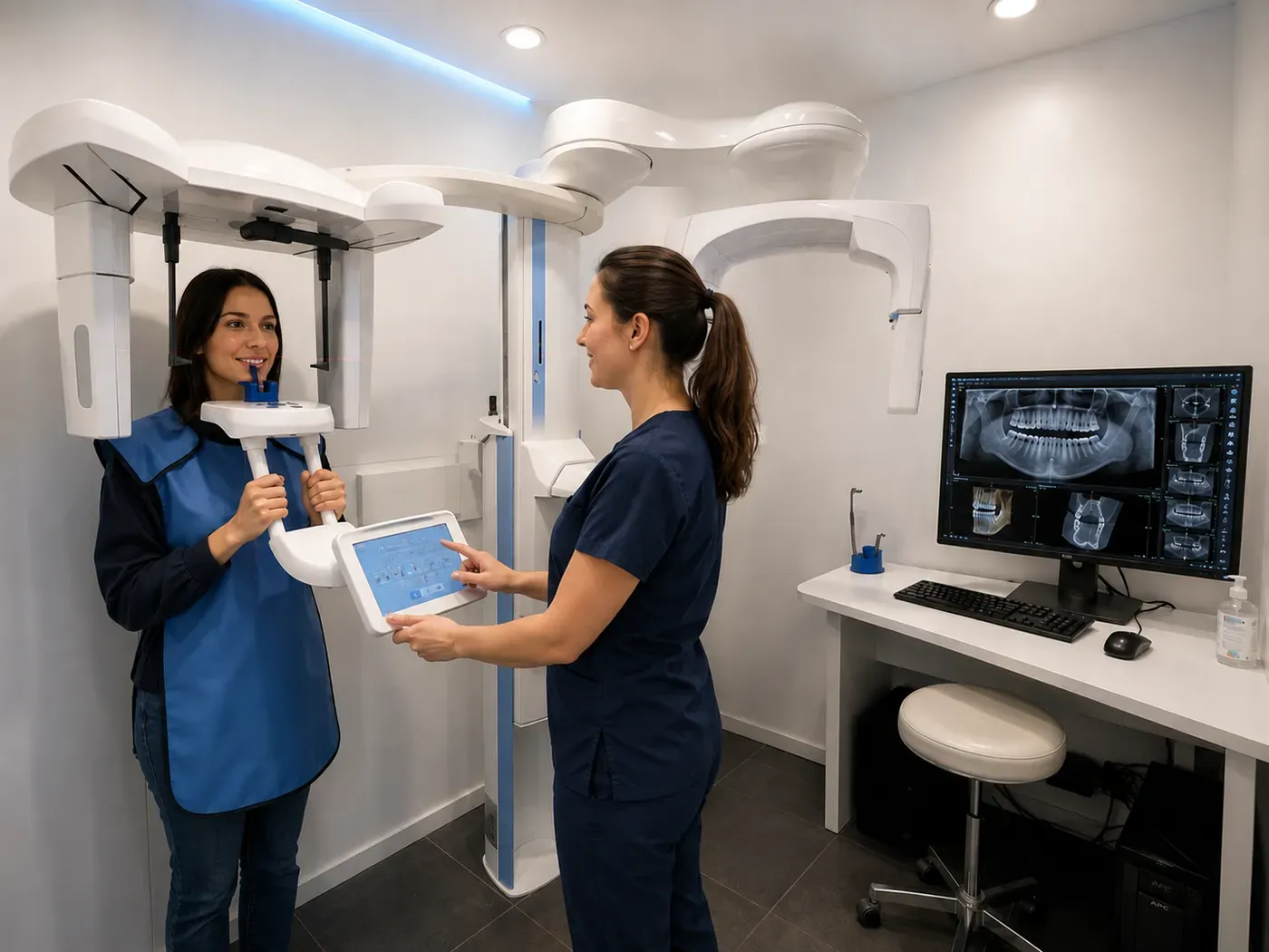
What is the examination you have been referred for? Here is what each one shows, and why.
A three-dimensional image of your teeth and jaw, to pinpoint to the millimetre what a conventional X-ray does not show. It is useful before an implant or a complex root canal treatment. Painless, in a few seconds.
An overall view of the whole mouth in a single image, to spot hidden decay, a wisdom tooth or a bone problem before it gets worse.
A side-view X-ray that measures the position of your jaws and teeth precisely. It is the basis of orthodontic diagnosis.
In practice
Four steps, from referral to clinical report. The examination itself takes a few seconds and requires no particular preparation.
Your dentist, orthodontist or surgeon requests the examination, specifying the area to be examined and the clinical indication. That precision makes it possible to limit the field of view to what is strictly necessary.
Book online through Doctena or by telephone on (+352) 26 68 11 30 1. Bring the referral with you and, if you have them, your previous examinations: they serve as a point of comparison.
Painless, with no injection and no particular preparation. You stay still for a few seconds while the unit rotates around your head. Jewellery and removable dentures are taken out beforehand.
The images are interpreted and then sent to the referring practitioner within 24 to 48 hours, together with the written clinical report. There is nothing for you to do: the transfer is direct.
The examination referred depends on the clinical question being asked. Here are the most common indications.
Yes. All examinations at the Cabinet d’Imagerie Dentaire are carried out by referral only, from a dentist, an orthodontist or a surgeon. The referral specifies the area to be examined and the clinical indication, which makes it possible to limit the examination to what is strictly necessary. The clinical report is sent to the referring practitioner within 24 to 48 hours of the examination.
The panoramic X-ray gives an overall two-dimensional view of the mouth: hidden decay, wisdom teeth, the condition of the bone. The lateral cephalogram is a side-view X-ray that measures the position of the jaws: it is the basis of orthodontic diagnosis. The Cone Beam, or CBCT, produces a three-dimensional volume, useful before an implant, a complex extraction or a difficult root canal treatment.
Every radiological examination involves a dose of X-rays. Our equipment is digital and the field of view is limited to the area named on the referral, which reduces the dose delivered compared with a wide-field examination. That is precisely why these examinations are carried out by referral only, with a justified clinical indication. The examination itself is painless and takes a few seconds.
Imaging most often supports orthodontic or endodontic treatment. The facilities are at the Luxembourg-Merl clinic.
Referring dentists
You refer, we carry out and interpret, you receive. The patient has nothing to forward, and you have nothing to chase.
3D volume or digital images, sent in DICOM format or through a secure viewing link, usable in your planning software.
The interpretation of the images in the light of the clinical indication given on the referral, within 24 to 48 working hours.
Where previous examinations are supplied by the patient or by you, any change is noted in the clinical report.
A question about an examination already carried out is settled by telephone with the practitioner who interpreted it, without needing a new referral.